
Chest Injuries
Warning
Objectives
To guide the management of patients presenting with chest injuries.
Scope
This guideline describes a standard approach and core management principles for patients with chest injuries.
It does not describe specific procedural details as these are contained within CGO procedure guides which will be linked where relevant.
It does not describe speciality surgical or intensive care management beyond the Emergency Department.
Audience
This guideline is intended for the use of registered healthcare professionals fulfilling a general role in a forward medical location or in an Emergency Department on deployed operations.
Initial Assessment & Management
PrinciplesAssessment and management of chest injuries should take place as part of the structured assessment of the trauma patient following standard principles (e.g. the MARCH assessment protocol).
Management should be focussed on maximising oxygen delivery and avoiding the ventilatory component of cellular hypoxia and shock.
Obstructive shock must be corrected as soon as possible following traumatic chest injury.
Chest injuries are extremely painful. Early adequate analgesia, as well as being humanitarian, will facilitate ventilation and oxygenation reducing later complications. See Defence Analgesia CGO for more details.
Where it is available, AVOID the use of Entonox for patients with chest injuries as there is a risk of increasing pneumothorax volume with its use. Penthrox (Methoxyflurane) is safe for patients with chest injuries.
Chest injuries continue to evolve throughout the early phase of care and many classically described features of established conditions may not initially exist.
Understanding the mechanism of injury is vital to assessing the likely pattern of pathology.
Meticulous physical examination should be conducted, again using a structured approach (e.g. the TCCC(UK) WORSEN-FLAP assessment) to identify conditions. This should be repeated with any change in the patients condition to detect evolving pathology.
This assessment must include key concealed areas including the neck, back and axillae.
Patient positioning is extremely important in chest injury management and where possible (e.g. when spinal injuries are not a significant concern) patients should be enabled to sit up or in a position of comfort to facilitate effective ventilation.
LIFE THREATENING CONDITIONSThe following conditions are generally considered life threatening in the initial treatment phase and require appropriate early management:
Blast Lung:
- Delayed presentation (24-48hrs) following blast wave exposure.
- Typical presentation with progressive cough, dyspnoea, haemoptysis, tachypnoea and hypoxia.
- Initial management should focus on exclusion of other occult injuries, oxygenation and strict fluid balance.
- Require urgent evacuation as high risk of progressive ventilatory failure and need for ventilatory support.
Open Pneumothorax:
- Penetrating thoracic injury creating a tract between the pleural cavity and the atmosphere. Large defects may lead to progressive air influx, lung collapse and ventilatory failures.
- Typical presentation is with an open chest wound and respiratory distress. May or may not have the classically described "bubbling" or "sucking" sounds.
- Management as per the Open Pneumothorax CGO.
- Require urgent evacuation as high risk of progressive ventilatory failure and need for ventilatory support.
Tension Pneumothorax:
- Occurs in blunt or penetrating trauma where a unidirectional air leak develops into the pleural space leading to progressive air accumulation, lung collapse and ventilatory failure.
- Rising intra-pleural pressure may also cause mediastinal shift and decreased venous return leading to obstructive shock.
- In the spontaneously ventilating patient this commonly presents with ventilatory failure. Conversely, in positive pressure ventilation, obstructive shock may develop after only a few breaths.
- Management is with decompression, either via Needle Decompression or if skills exist and in line with the Thoracostomy CGO with open (finger) thoracostomy.
- Require urgent evacuation as high risk of progressive ventilatory failure and need for ventilatory support.
Massive Haemothorax:
- Injuries to the thoracic wall vessels, lung (including hilum), cardiac injuries and/or thoracic vessels may all cause major haemorrhage into the pleural cavity.
- Predominantly leads to the development of hypovolaemic shock with worsening ventilatory failure in extreme cases.
- Management is in line with the Initial Management of the Shocked Patient CGO including damage control resuscitation measures.
- A chest drain (tube thoracostomy) may be required in line with the CGO but prioritise filling (blood product resuscitation) over draining.
- Require urgent evacuation as high risk of progressive shock and need for emergency transfusion and surgical management.
Cardiac Tamponade:
- Penetrating (or rarely blunt) trauma to the heart causing direct cardiac or coronary vessel injury with blood loss into the pericardium and progressive increase in pericardial pressure. Progressive pressure rise leads to obstructive shock and rapid cardiac arrest.
- Pre-cordial penetrating injury and shock should prompt consideration of the diagnosis. "Beck's Triad" of muffled heart sounds, hypotension and distended neck veins is unreliable especially where there is concurrent hypovolaemia.
- Point Of Care Ultrasound (POCUS) may aid the diagnosis where this is available, especially in blunt trauma.
- Management is with rapid evacuation to surgical care, if possible with the use of enhanced pre-hospital critical care assets capable of delivering Resuscitative Thoracotomy (See Traumatic Cardiac Arrest CGO).
Flail Chest:
- Two or more adjacent ribs fractured in two or more places leading to loss of chest wall integrity with a "flail" segment of chest wall able to move paradoxically with breathing.
- Flail segments may be visible or palpable on chest examination. May be difficult to detect if the injury is "splinted" by chest wall muscles, involves predominantly posterior ribs or in symmetrical/bilateral injuries. Concealed injury initially may be exposed in the tiring patient.
- Initial management should focus on exclusion of other occult injuries, oxygenation and effective analgesia. There is no benefit in attempting to "splint" the chest wall with improvised measures.
- Require urgent evacuation as high risk of progressive ventilatory failure and need for ventilatory support.
Great vessel injury:
- May occur with penetrating injuries or in blunt trauma involving rapid deceleration forces.
- At the extreme end presents with cardiac arrest or profound shock. Moderate aortic injuries may present more like a Spontaneous Aortic Dissection (Acute Aortic Syndrome). Injuries may also be asymptomatic initially.
- Management is with early evacuation for advanced imaging and surgical care.
Other chest injuriesThe following chest injuries are not immediately life threatening but require careful management and decision making around evacuation:
Rib fractures:
- Simple rib fractures are extremely common in chest trauma. Consideration should always be given to underlying injuries (e.g. haemo/pneumothorax).
- Specific additional considerations include the risk of major vessel injury with fractures of ribs 1-3 and liver, splenic and renal injuries with lower rib fractures (Junctional Injury).
- Management is focussed on effective analgesia, oxygenation and avoidance of complications.
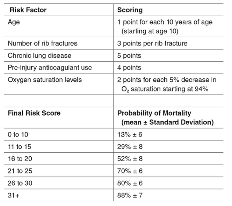
- The "STUMBL" score may be used to guide management including urgency of evacuation, analgesia required and likelihood of complications (see accordion content).
- Most suspected rib fractures will require evacuation unless no complications are suspected, analgesic requirements are minimal and patients role is non-dynamic OR the tactical situation dictates otherwise.
- If patients with simple rib fractures are not being evacuated they must have adequate analgesia, be given chest physiotherapy/injury advice and have regular review by the deployed medical team.
Sternal fractures:
- Suspicion of sternal fracture raises the possibility of blunt cardiac injury, particularly where there is clinical evidence of displacement of a fracture.
- A 12 lead ECG should be completed where possible.
- Point of Care Troponin tests, if available, can also be used to help with risk stratification.
- Management is focussed on effective analgesia, oxygenation and avoidance of complications.
- In the absence of a normal ECG and negative point of care troponin, all suspected sternal fractures should be evacuated for exclusion of cardiac injury, unless the tactical situation dictates otherwise.
- If 12 lead ECG and point of care troponin testing are normal AND no other complications are suspected, analgesic requirements are minimal and patients role is non-dynamic clinicians may consider not evacuating and management in line with simple rib fracture management above.
Simple pneumothorax or haemothorax:
- More common than tension pneumothorax in both blunt and penetrating trauma, commonly co-exist.
- Clinical signs and symptoms vary by volume with small injuries causing minimal findings but progressive respiratory distress and/or hypovolaemia with increased size.
- POCUS may aide diagnosis where this is available.
- Management is focussed on effective analgesia, oxygenation and monitoring for deterioration.
- Tube Thoracostomy (surgical chest drain) is rarely required in initial management but indications can be found in the Thoracostomy CGO.
- Require evacuation as risk of progressive ventilatory failure or continued haemorrhage and shock.
Advanced Assessment & Management
Principles
Advanced assessment and management of chest injuries follows the same core principles as initial management.
Serial assessment and examination adds value in the context of evolving disease after chest trauma.
Meticulous attention to detail around oxygenation, analgesia and the early detection of progressive ventilatory failure is vital.
The approach to analgesia should be scalable and multi-modal in line with Defence Analgesia CGO . Example options include:
- Patient positioning and support
- Simple analgesic agents - Paracetamol, NSAIDS
- Opiate analgesic agents - morphine, fentanyl, oxycodone (bolus, infusion or PCA)
- Ketamine (bolus, infusion or PCA)
- Topical local anaesthetic agents (e.g. Lidocaine patch)
- Regional anaesthesia (e.g. Erector Spinae Block, Serratus Anterior Plane Block)
Investigations
The approach to investigating chest injuries in the deployed setting will vary depending on modalities available.
The gold standard remains contrast enhanced CT thorax which will enable rapid and accurate identification of major thoracic injuries including more occult patterns such as great vessel injury within the thoracic cavity. Chest injuries in both penetrating and blunt trauma are rarely isolated and there should be a very low threshold to extending this to a full CT Trauma Series.
Where this is not immediately available a combination of Chest X-Ray with Point of Care Ultrasound (POCUS) can be highly effective in combination with clinical examination, particularly if these are performed serially with any change in clinical condition.
A standard panel of trauma blood tests should be taken where possible to include as a minimum a full blood count, coagulation screen and group and save/crossmatch sample. Viscoelastic assays (such as TEG/ROTEM) should be used if available in keeping with the Major Haemorrhage CGO. Serum troponin testing may be useful in the identification or exclusion of sub-clinical blunt cardiac injury.
LIFE THREATENING CONDITIONS
Rapid advanced management of the following conditions after the initial phases detail above may further reduce morbidity and mortality:
Blast Lung:
- Careful attention to oxygenation and fluid balance may prevent acute deterioration.
- High risk of requiring intubation and positive pressure ventilation, seek ICU support.
Open Pneumothorax:
- Likely to require surgical chest drain placement in line with Thoracostomy CGO.
- Surgical closure options should be reviewed with deployed surgical teams +/- reach back for cardiothoracic opinion.
Tension Pneumothorax:
- Likely to require surgical chest drain placement in line with Thoracostomy CGO.
- High air output may indicate broncho-pleural fistula and should be reviewed with deployed surgical teams +/- reach back for cardiothoracic opinion.
Massive Haemothorax:
- Likely to require surgical chest drain placement in line with Thoracostomy CGO.
- Blood transfusion and associated management should be instituted in line with the Major Haemorrhage CGO <LINK>
- High blood output from a chest drain of >1000mls immediately or 200mls/hr should prompt urgent surgical review with deployed surgical teams +/- reach back for cardiothoracic opinion.
Cardiac Tamponade:
- In cardiac arrest should proceed as per the Traumatic Cardiac Arrest (TCA) CGO.
- If identified in a peri-arrest patient should prompt urgent surgical management with thoracotomy and pericardial decompression.
- Judicious blood transfusion may temporise an evolving cardiac tamponade allowing transfer to theatre for formal thoracotomy.
Flail Chest:
- Careful attention to analgesia and oxygenation may prevent acute deterioration
- High risk of requiring intubation and positive pressure ventilation, seek ICU support.
- Rib fixation may be required for multiple rib fractures or flail chest but would not be completed in a deployed hospital setting.
Great vessel injury:
- If identified, deployed management is supportive including careful ICU control of blood pressure and heart rate to avoid worsening vessel injury.
- Expedient evacuation should be sought for surgical or percutaneous vascular intervention.
Other chest injuries
Attention to detail in the management of the following less immediately life threatening conditions can prevent deterioration and subsequent morbidity/mortality:
Rib fractures:
- Management continues to be focussed on effective analgesia, oxygenation and monitoring for development of complications.
- The "STUMBL" score may be used to guide management including care environment, urgency of evacuation, analgesia requirements and likelihood of complications (see below).
- Early physiotherapy is vital in the avoidance of complications and should be instituted as soon as practicable in the deployed setting (See below for physiotherapy advice).
- Rib fixation may be required for multiple rib fractures or flail chest but would not be completed in a deployed hospital setting.
Sternal fractures:
- A lateral sternal X-Ray should be conducted if CT Thorax is not available or felt not to be required but the suspicion of a sternal fracture exists.
- All patients should have a 12 lead ECG and High Sensitivity Troponin blood tests.
- POCUS may be useful in excluding a pericardial effusion or evolving tamponade. In more advanced hands this may also identify regional wall motion abnormalities secondary to cardiac contusion.
- Management continues to be focussed on effective analgesia, oxygenation and monitoring for development of complications.
- In the absence of cardiac contusion
- Fixation is rarely required, only in highly unstable or severely displaced injury and would not be completed in a deployed hospital setting.
Simple pneumothorax or haemothorax:
- Principles are similar to in the initial phase.
- Clinical signs and symptoms vary by volume with small injuries causing minimal findings but progressive respiratory distress and/or hypovolaemia with increased size.
- Chest X-Ray combined with POCUS should be used to confirm diagnosis and inform decision making around volume in the absence of CT Thorax.
- Tube Thoracostomy (surgical chest drain) may be required and indications can be found in the Thoracostomy CGO. Discussion with the deployed surgical team and/or reach back for expert advice is important as although historically even very small haemo/pneumothoraces were drained, modern trends are towards conservative management unless compromised.
Blunt cardiac injury:
- Spectrum of conditions from minor ECG/Troponin changes to major septal, free wall or valvular injuries.
- Diagnosis via ECG, High Sensitivity Troponin blood tests and POCUS.
- Minor injuries including ECG changes, and troponin rises without injury on POCUS should have a 24-48hr period of observation and serial ECG monitoring.
- Major injuries will likely present with significant haemodynamic instability and should be treated in consultation with the deployed surgical teams +/- reach back for cardiothoracic opinion.
Pulmonary Contusion:
- Injuries commonly evolve over the 24-48hr period post thoracic trauma with gradual reduction in lung compliance and evolution of respiratory distress.
- When pulmonary contusions are identified on X-Ray or CT patients should have close monitoring for deterioration.
- Careful attention to oxygenation and fluid balance may prevent acute deterioration.
- High risk of requiring intubation and positive pressure ventilation, seek ICU support.
Ruptured diaphragm:
- Can occur in both blunt and penetrating injury to the thorax. In blunt trauma this is most commonly with side impacts (e.g. T-Bone road traffic collision).
- May be initially asymptomatic, or may have reduced air entry to lower zone of chest, respiratory failure or occasionally acute abdomen secondary to strangulation of bowel loops through the hernia.
- May be identified on Chest X-Ray, requires CT imaging to definitively confirm the extent and site of the injury.
- Most will require surgical repair, although not necessarily in a deployed hospital setting, and should be treated in consultation with the deployed surgical teams +/- reach back for cardiothoracic opinion.
Oesophageal injury:
- Uncommon, the oesophagus is generally well protected in the posterior mediastinum, May occur in penetrating trauma, crush injury or blows to the epigastrium.
- Leaks present with pain out of proportion to presentation, particularly on swallowing. May evolve rapidly into septic shock secondary to mediastinitis. May display surgical emphysema.
- Chest Xray may demonstrate left sided pneumothorax, effusion or pneumo-mediastinum. Particularly when these present without obvious rib fractures.
- Management should be in consultation with the deployed surgical teams +/- reach back for cardiothoracic opinion.
Prolonged Casualty Care
Patients with significant chest injuries are at high risk of deterioration, and every effort should be made to prioritise evacuation where this is practicable.
Deteriorations are most commonly secondary to the following conditions and PCC management should focus on reducing the risks of these where possible as well as identifying and managing them early when they are occurring:
- Evolving blast lung injury
- Evolving pulmonary contusions
- Increasing volume of haemothorax
- Increasing volume of pneumothorax (including evolving tension)
- Progressive ventilatory failure secondary to pain and/or exhaustion
- Infection (including from battlefield injury, iatrogenic after procedures and pneumonia due to hypoventilation/atelectasis)
- Complications of invasive procedures
Management:
Where possible allow patient to adopt a position to optimise ventilation, usually as upright as possible. If a patient must be kept flat and there are unilateral injuries consider lateral positioning with the injured side downward.
Monitor observations as closely as possible including respiratory rate, heart rate, oxygen saturations, blood pressure and temperature.
Deteriorating observations should prompt full re-assessment in line with the initial principles described above and management of any conditions identified.
Maintain oxygen saturations of 94-98% where oxygen supplies allow. If oxygen supplies are limited it is preferable to accept a lower target (e.g. 88-92%) for a longer period rather than attempt to sustain higher saturations before exhausting supplies and having severe acute hypoxia as a result.
Maintain careful management of analgesia, target the ability to take deep breaths and cough without significant pain.
Where possible begin chest physiotherapy exercises as detailed in the accordion content. Encourage mobilisation of the patient wherever possible.
Avoid smoking as this may increase the risk of chest infection.
If there is an open chest injury, or an invasive procedure has been performed, commence antibiotics in line with deployed antimicrobial guidance
Monitor for signs of hypoventilation/atelectasis associated infection, which will present in a similar way to an evolving pneumonia. Have a low threshold to start antibiotics in line with deployed antimicrobial guidance.
Use intravenous fluids cautiously in patients at risk of blast lung injury or pulmonary contusion. Excessive use may worsen these conditions.
Continue to monitor and manage surgical chest drains in line with the Thoracostomy CGO. Be diligent to avoid drain kinking or obstruction which may precipitate rapid tension pathology.
Ongoing drain blood loss should be managed in line with the Major Haemorrhage CGO. There is no value in clamping drain output in a false attempt to "tamponade" bleeding.
Paediatric Considerations
Assessment
The differences in anatomy and physiology of paediatric trauma patients alter injury patterns, assessment strategies and management approaches to chest trauma in paediatric patients. Most children older than 13 years of age are however grossly similar to an adult of similar stature.
The principles outlined in the initial and advanced assessment sections are common across all age groups.
Meticulous physical examination should be conducted, using a structured approach (e.g. the TCCC(UK) WORSEN-FLAP assessment) to identify conditions. This should be repeated with any change in the patients condition to detect evolving pathology.
External signs of chest injury may be reduced in paediatric patients, in particular children's ribs are highly flexible and underlying injuries may be significant without fractures. Conversely if rib fractures are seen this indicates a high degree of energy transfer and suspicion of underlying injuries should be similarly high.
Investigation strategies in paediatric thoracic trauma do differ from adults. In particular the "ALARA" (As Low As Reasonably Achievable" principles around ionising radiation limit the use of Thoracic CT imaging.
The following strategy is recommended:
- Chest X-Ray is the primary imaging modality for blunt paediatric trauma. In a conscious, stable child with a blunt mechanism and a normal Chest X-Ray, CT thorax is almost never required.
- CT thorax should be used first line, where this is available, for penetrating chest trauma, in poly-trauma or shocked patients, and where there is severe distracting injury or brain injury.
- Where CT is not immediately available POCUS can be useful in identifying thoracic injuries.
ManagementManagement should be focussed on maximising oxygen delivery and avoiding the ventilatory component of cellular hypoxia and shock. This is particularly true for children where low functional residual capacity and high basal metabolic rates may lead to rapid desaturation.
Chest injuries are extremely painful, and children are even more likely to hypoventilate due to pain than adults. Early adequate analgesia, as well as being humanitarian, will facilitate ventilation and oxygenation reducing later complications. See Defence Analgesia CGO for more details.
The principles of management of most traumatic chest injuries are the same as for adults. The following modifications should be considered:
- There may be a lower clinical threshold to intubate and ventilate children to facilitate thoracic procedures and prevent severe respiratory distress.
- Supported ventilation without a definitive airway will likely lead to gastric distension and diaphragmatic splinting. Early decompression with a naso/orogastric tube is recommended.
- Needle decompression and surgical chest drain placement will require alterations in techniques and equipment. See relevant CGOs for guidance.
- Chest seals may need to be cut down in size for use.
- Chest drains placed for haemothorax should be monitored closely. Drain output of >3mls/kg/hr should be considered high output and require urgent surgical review.
Stage 1:
Sit upright and mobilise as early as possible, use analgesia effectively to facilitate this and the following exercises:
Deep Breathing:
Deep breaths are a simple way to fill your lungs up with air and clear any phlegm that builds up in your lungs. Practice the following exercises hourly throughout the day.
Start in a comfortable position, ideally sitting upright in the bed or chair with your shoulders relaxed.
1) Take a slow, deep breath in through your nose to fill up the bottom of your lungs
2) Hold this breath for a count of 3 (if you are able)
3) Breathe out slowly through your mouth.
4) Repeat steps 1-4 a total of 10 times (don’t do more as you may get dizzy)
5) Repeat every hour during the day when you are awake.
Supported cough:
Coughing is the normal way to clear phlegm from your lungs. Following your deep breathing, you should perform a cough.
When you need to cough, use a pillow or rolled up towel to support the area of your chest wall that is uncomfortable to help reduce the pain.
Huffing:
Twice an hour you should take a deep breath in, and then force your breath out quickly- as if you were steaming up a mirror before wiping it clean - making a ‘huffing’ sound.
This technique will aid sputum clearance and help to reduce the risk of complications.
Stage 2:
If able to conduct the above without significant discomfort and mobilising after analgesia progress to the following exercises:
Rotation:
Sit up straight, with your arms across your chest.
Keeping your legs still, gently rotate to one side as far as you can go without pain and provided you feel no more than a mild to moderate stretch.
Hold for 1 – 2 seconds and repeat 10 times to each side
Shoulder Blade Squeezes:
Begin sitting or standing tall with your back straight.
Squeeze your shoulder blades together as far as you can go without pain and provided you feel no more than a mild to moderate stretch.
Hold for 1-2 seconds and repeat 10 times.