Objectives
To guide the immediate management of patients with suspected cervical spine injuries including clearing the cervical spine.
Scope
This guideline describes the immediate management of patients who may have a suspected cervical spine injury based on mechanism of injury and clinical findings, and provides guidance for prolonged casualty care of these patients if required. It does not include management or interventions that might be considered by a specialist.
Audience
This guideline is intended for the use of registered healthcare professionals fulfilling a general role in a forward medical location or in an Emergency Department on deployed operations.
Initial Assessment & Management
Consider mechanism of injury
Examples of high risk mechanisms include:
Blast injury where a casualty has been projected or thrown by the blast
Fall from a height greater than 1 metre
Motor vehicle collision at high speed, in which the vehicle has rolled or from which the casualty has been partly or fully ejected
Accidents involving quad bikes
Injury with axial load to the head – for example falling headlong or diving into shallow water
Unstable cervical spine injuries are very rare in the context of isolated penetrating neck injuries.
Non-permissive environment
Care should be taken to minimise head and neck movement if there is a high risk mechanism for cervical spine injury BUT control of major haemorrhage and airway opening manoeuvres must not be compromised.
Evacuation to a more permissive environment takes precedence over spinal immobilisation. Collars, scoop stretchers and head blocks should not be used in this phase of care.
Semi-permissive environment
If there is a high risk mechanism for cervical spine injury or the patient complains of neck pain, then restrict movement of the cervical spine initially by manual in-line stabilisation and then the application of head blocks (improvised or bespoke) and tape.
A cervical collar should be used only for the extrication of an unconscious casualty who has been subjected to a high risk mechanism for cervical spine injury. Once extrication is complete, the collar should be removed; head blocks and tape must remain in place for onward transfer.
Do not use a cervical collar if there is a penetrating neck injury present. A collar may mask wounds and hide evolving haematoma development.
Do not attempt manual in-line stabilisation or other immobilisation in a patient who is agitated and combative - pinning the head down while the body moves around only risks exacerbating any injury. Reassure and calm the patient, giving analgesia and sedation as needed until the patient is sufficiently compliant to tolerate safe immobilisation.
Permissive environment
Initially consider whether the cervical spine can be cleared using the Canadian C-spine rule.
If the patient has paraesthesia, imaging is required (NEXUS criteria will also not be met).
If there are any high risk factors, the Canadian C-spine rule cannot be applied.
Otherwise, if there are any low risk factors present, proceed to active neck movement.
If the patient can actively rotate their neck by at least 45° both left and right, there is no need for imaging.
The Canadian C-spine rule can only be used if there are no high risk factors and if there is at least one low risk factor present. If the Canadian C-spine rule cannot be used then consider whether the NEXUS criteria can be used.
No altered level of consciousness or intoxication
Head blocks and tape - if applied correctly may remain in situ for several hours with minimal discomfort, although improvised head blocks may be more uncomfortable and often obstruct hearing.
Scoop stretcher and straps - represents standard civilian practice but avoid using for prolonged periods over 60 minutes due to the risk of pressure sores.
Vacuum mattress - may be useful for prolonged evacuation and can be helpful at splinting other injuries.
Cervical collar - should only be used for extrication in patients who are unconscious. Collars should not be used for evacuation/transfer but should be removed when extrication is complete and the patient has been placed on a scoop, vacuum mattress or similar. Do not use a collar if there is penetrating neck injury.
Examination of the Back and Log Rolling
In cases of blunt force trauma where there is no concern about a possible penetrating injury to the back, formal examination of the back may be deferred while the patient is transferred to a facility with imaging capabilities. Examination of the back in a forward location in blunt force is only required if there is bleeding or other evidence of a wound, or as part of an attempt to clear the spine of injury in patient without other significant injuries who might be able to mobilise and avoid evacuation. However, log rolling may be needed to allow removal of clothing/body armour and/or repositioning of the patient.
In penetrating trauma, by contrast, early, thorough examination of the back is essential, and must at the latest be completed by the end of the Respiration phase of MARCH assessment. However, the incidence of spinal injury in penetrating trauma is very low: a conscious patient may be able to sit up to allow the back to be examined. Careful patient handling remains important to avoid disrupting clot formation and precipitating worsening haemorrhage, but log rolling is not needed.
In blast injury there is both a high risk of penetrating injury to the back and a high risk of spinal injury if the patient has been projected or thrown. Early, thorough examination of the back is essential and must at the latest be completed by the end of the Respiration phase of MARCH assessment. If there are sufficient personnel to allow a log roll then this minimises the risk of disrupting a potential spinal injury, but if additional personnel are not available then do not delay examining the back to await a formal log roll. Consider the patient's cervical spine and roll as carefully and gently as possible with the personnel available to allow full exposure of the back.
Neurological Examination
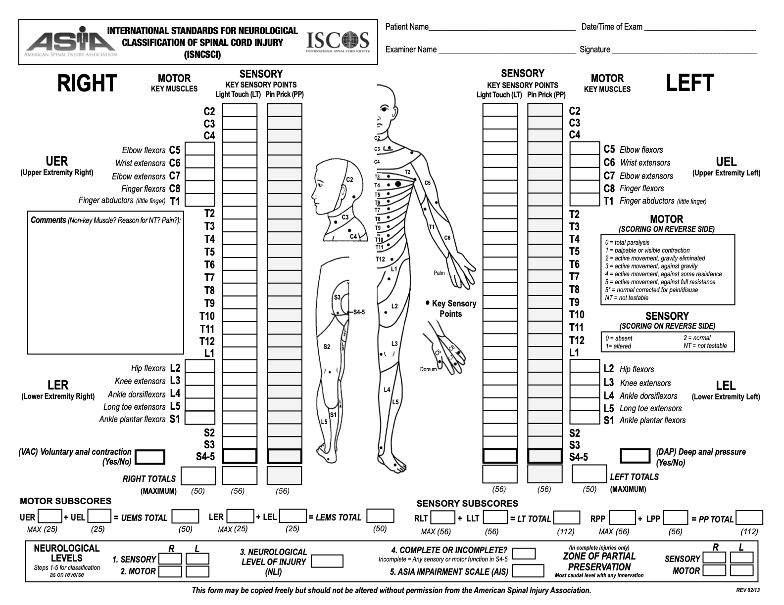
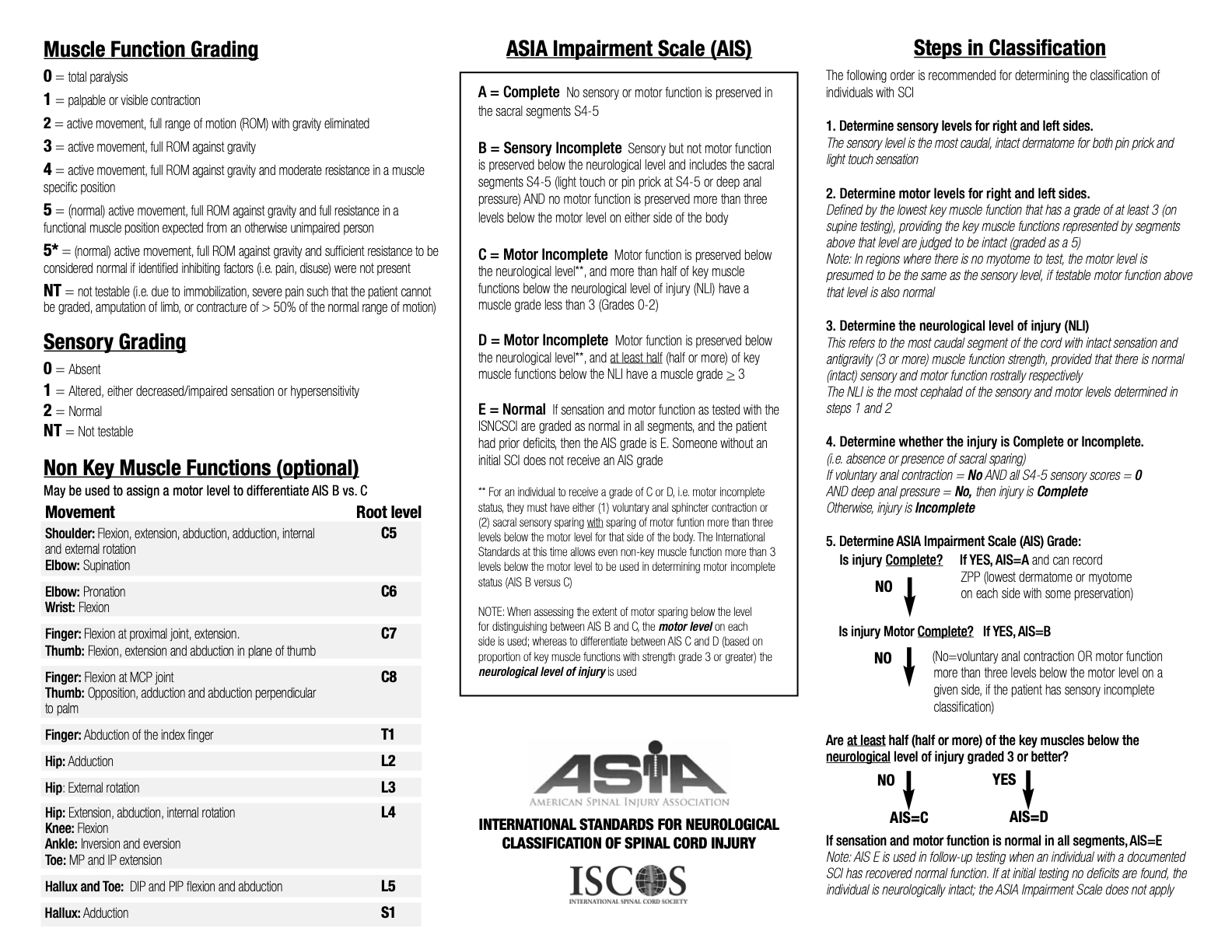
In conscious patients with suspected cervical spinal injury, initial gross neurological examination should be followed by a full neurological assessment as soon as circumstances permit. Neither imaging nor evacuation should be delayed to allow this to be undertaken, but if a casualty is being held pending transfer then completing a detailed assessment that can be passed on to the specialist clinical team may support prognostication and planning. The ASIA chart (see accordion section below) is a useful tool to both guide and document neurological examination.
Advanced Assessment & Management
Airway Management
Patients with an injury above level of C4 are likely to have severe respiratory failure due to loss of diaphragm and intercostal muscle innervation, and will usually have initial airway management and ventilatory support delivered as part of immediate interventions under MARCH principles.
Patients with an injury between C4 and C8 are likely to retain some ventilatory function, but the loss of intercostal muscle function reduces functional vital capacity (FVC) and tidal volume by up to 60% and casualties are likely to tire quickly, so early intubation and mechanical ventilation are indicated.
Circulatory Support
In patients with a suspected spinal cord injury, fluid resuscitation should initially be used to target a mean arterial pressure (MAP) >85 mmHg to ensure spinal cord perfusion and reduce the risk of secondary injury. Vasopressor support may be required to achieve this - see separate guidance (link to follow).
If neurogenic shock is identified or suspected, follow the guidance in the CGO on spinal cord injury and neurogenic shock (link to follow).
Imaging
If the cervical spine cannot be cleared clinically then immobilisation should be used to protect the patient during evacuation to a facility equipped with imaging.
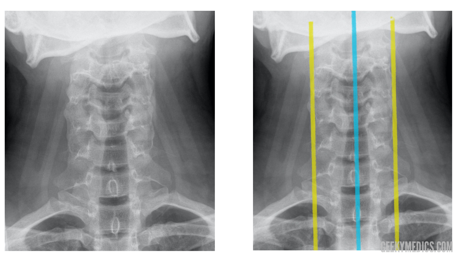
CT imaging is now generally regarded as the standard modality within UK practice for cervical spine imaging, but may not be available in the operational environment.
Plain film x-ray imaging should not be used to attempt to radiologically exclude cervical spine injury:
- if CT is readily available
- if the patient is already being evacuated to a level of care equipped with CT for other reasons
- if there is any evidence of a focal neurological deficit
- if the patient is unconscious
If attempting to use plain film x-ray imaging to radiologically exclude cervical spine injury, decision-making should be supported by reach back discussion if possible.
MRI imaging is required if a patient with a suspected spinal injury has a neurological deficit (motor or sensory deficit on examination), even there is no bony injury apparent on CT imaging - CT does not exclude significant ligamentous injury. Such patients will require evacuation to a level of care with MRI available, and should remain immobilised until specialist assessment. The ASIA chart (see accordion section) may be helpful to precisely document neurological examination findings.
Medication
Analgesia in acute spinal cord injury should follow the principles described in the analgesia CGO.
Based on current evidence, the early use of methylprednisolone, nimodipine and/or naloxone for acute spinal cord injury is not recommended.
Prolonged Casualty Care
Urgent evacuation is important as significant resources are required to deliver an appropriate level of care to patients with suspected or proven cervical spine injury, and delay may be associated with a risk of neurological deterioration and the development of secondary injuries including pressure sores.
In addition, patients with suspected, but ultimately excluded, cervical spine injury can be returned to normal duties most quickly by rapid evacuation to a facility with sufficient imaging capabilities to rule out significant injury.
If prompt evacuation is not possible then prolonged care of the casualty with suspected spinal injury should follow standard principles. In particular:
Monitor ventilatory requirements and consider the need for supplemental oxygen and/or ventilatory support.
Ensure that the patient is as comfortable as possible, and minimise the risk of pressure sores in both conscious and unconscious patients:
- Spinal boards and rigid cervical collars should only be used for extrication: both should be removed once extrication is complete. Immobilisation of the cervical spine with blocks and tape is sufficient.
- Scoop stretchers may be used for up to 60 minutes; if holding a casualty for longer than this then remove the scoop so that the patient is lying on a mattress or the most comfortable surface available.
- If a patient is fully alert and co-operative, they may be able to simply lie still without the need for the discomfort of full immobilisation.
- Reposition and log roll patients regularly (at least every four hours if possible) to avoid pressure sores.
Insert a urinary catheter; urinary retention is a frequent cause of agitation and physiological distress in patients with a suspected or confirmed cervical spine injury.
Continue to reassess symptoms for resolution or progression. Remember that it may become possible to clinically clear a patient's cervical spine, for example if intoxication resolves.
Paediatric Considerations
The management of children with a suspected spinal cord injury is challenging in the deployed environment as there is a much higher incidence of spinal cord injury without radiological abnormality (SCIWORA) than in adult patients. Consequently MRI is the accepted standard for imaging in children but this may not be available on operations.
The Canadian C-spine rule is only validated in adults, but the NEXUS criteria can be used to support the assessment of children.
If imaging is required:
- Consider possible availability of MRI in local civilian / partner force facilities
- Seek to evacuate for MRI if any neurological deficit
- Otherwise use CT if there is no access to MRI
- Plain film imaging is unlikely to be of value and should only be used when there is no means of accessing CT. Reach back discussion is recommended to support decision-making.
Remember that in all patients, but especially in children, normal CT imaging does not exclude spinal cord injury. Assess neurological function as thoroughly as possible given the age of the child and the circumstances of the assessment; manage unconscious casualties with caution until clinical assessment of neurological function is possible