
Acute limb ischaemia
Objectives
To guide the immediate investigation and emergency management of patients presenting to medical facilities with suspected Acute Limb Ischaemia (ALI).
Scope
These guidelines describe the immediate assessment and emergency management for patients with Acute Limb Ischaemia.
1. Features of Acute Limb Ischaemia in a Traumatic and Non-Traumatic context
2. Decision making around Acute Limb Ischaemia and patients presenting with the High and Tight Tourniquet.
3. Intra-operative approach to management of Acute Limb Ischaemia
4. Guidance on assessment and management of Reperfusion injury
Not included in the guideline:
1. Long term guidance regarding follow up, further investigation, long term deployability/ occupational health considerations for patient who have had ALI
Audience
This guideline is intended for the use of registered healthcare professionals fulfilling a general role in a forward medical location or in an Emergency Department on deployed operations. (Role 1, 2-3 (ED, Ward)).
Initial Assessment & Management
Classification into traumatic and non-traumatic acute limb ischaemia
The aetiology of Acute Limb Ischaemia is important to consider from the outset as this dictates the management of the situation. The key categories for aetiology can be divided into Traumatic and Non-Traumatic.
| Traumatic ALI | Non-Traumatic ALI |
| Complete/ partial transection of artery |
Embolism from proximal source; Atrial/ ventricular thrombus Paradoxical embolism through PFO Fat embolus Aneurysmal disease Tumour Mycotic source |
| Dissection of artery | Vasculitis |
| Arterial thrombus or embolus from intimal trauma to vessel |
Thrombus in situ; Atherosclerosis Ulcer Plaque Aneurysm |
|
Extramural Occlusion cause; Applied Tourniquet/ Occlusive dressing/ packing Fracture Dislocation Haematoma Foreign body |
Dissection Acute Aortic syndrome |
| Compartment Syndrome | Low flow cardiac output states |
| Venous ischemia- Phlegmasia cerula dolens, alba dolens |
Classification into non-traumatic and traumatic is imperative as traumatic ALI is associated with bleeding which requires urgent damage control surgery, control of haemorrhage and restoration of arterial blood flow.
Major Vascular Injury and management of catastrophic haemorrhage is covered in TCCC and the Major Vascular Injury CGO.
ALI is defined as a sudden reduction in arterial perfusion that poses an immediate threat to limb viability (Acar et al., 2013; Björck et al., 2020). It is a vascular emergency requiring urgent assessment and management. ALI is distinguished from chronic limb ischaemia with a duration of 14 days or less.
ALI should be classified according to the Rutherford classification to guide urgency of intervention and likelihood of limb salvage (Rutherford et al., 1997) see table 1. Limbs classified as Rutherford IIa or IIb require urgent vascular intervention to restore blood flow and salvage the limb. Grade III are non-salvageable and require primary amputation or palliation.
| Grade | Category | Sensory loss | Motor deficit | Prognosis | Arterial Doppler signals | Venous Doppler signals |
| I | Viable | None | None | No immediate threat | Audible | Audible |
| IIA | Marginally threatened | None or minimal (toes) | None | Salvageable if promptly treated | Inaudible | Audible |
| IIB | Immediately threatened | More than toes | Mild/moderate | Salvageable if promptly revascularized | Inaudible | Audible |
| III | Irreversible | Profound, anaesthetic | Profound, paralysis (rigor∗) | Major tissue loss amputation. Permanent nerve damage inevitable | Inaudible | Inaudible |
If acute limb Ischaemia is suspected an urgent referral and evacuation to a medical treatment facility with 24-hour vascular surgery capability should be made as per deployed OPCP.
UK DMedC DHC surgical resource for investigating and managing vascular pathology is limited. Specifically, 6mm/8mm Dacron graft for shunting, limited suture size (5-0 double ended prolene) and no microvascular instruments.
Initial management should include prompt intravenous access, administration of systemic anticoagulation with unfractionated Heparin (UFH) if available. A bolus dose of 75 Units/Kg/IV is recommended (Björck et al., 2020). Alternatively, subcutaneous therapeutic low molecular heparin can be used.
Resuscitation/ correction of dehydration.
Oxygen should be administered and titrated to an Oxygen Saturation of >96%.
Urgent Vascular imaging should be sought where available, CTA in the first instance but Duplex Ultrasound or Magnetic Resonance Angiography can also be considered if expertise available. Administration of Heparin should NOT be delayed until after imaging if ALI is suspected.
Initiate a Heparin infusion, if possible. A 4 hourly APTTR is required to titrate dose. If long term infusion is not feasible then single sc Low Molecular Weight (LMWH), therapeutic dose can be considered with guidance from a vascular specialist.
Assessment of limb viability should be completed and should form the foundation of the Vascular referral.
Management of traumatic ALI
1. The below guideline describes management in the context of UK DMedC DHC
2. After initial assessment surgical revascularisation should be considered.
Time is tissue
Acute Limb Ischaemia is a surgical emergency with successful limb salvage dependant on time to reperfusion.
Early recognition and prompt restoration of arterial flow are essential to minimise ischaemic injury. Revascularisation may be achieved by temporary vascular shunt or definitive arterial repair.
Major haemorrhage is associated with significantly worse limb salvage and survival outcomes. Patients requiring massive transfusion have an increased risk of mortality and limb loss. In this context, tissue tolerance to ischaemia is reduced, and urgency of reperfusion heightened.
Optimal outcomes are associated with restoration of arterial flow as early as possible. In traumatic lower limb arterial injury, delayed revascularisation is associated with increased amputation rates.
In select cases, particularly in austere or deployed environment, limb sacrifice may be required to preserve life. This decision should be made early and involve senior clinical input within a multidisciplinary team (MDT).
Recognition and evacuation
Prompt recognition of ALI is essential to initiate immediate management and facilitate rapid evacuation to a vascular specialist service.
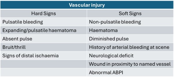
Clinical signs of vascular injury may be defined as hard or soft.
Hard signs of arterial injury suggest a high probability of significant arterial injury and require immediate surgical exploration.
Soft signs are suggestive but not indicative of arterial injury. They require urgent evaluation but do not mandate urgent surgery. A period of observation and repeat examination may be necessary.
For soft signs consider systemic anticoagulation if no contraindications, early reach back for vascular specialist advice and evacuation to an appropriate medical treatment facility
Certain orthopaedic injuries are strongly associated with underlying arterial trauma. If these bones are injured have a high index of suspicion for vascular injury, assess for signs of limb ischaemia.
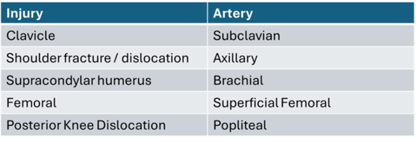
Vascular injury may be occult, even in the presence of palpable pulses. Serial clinical examinations are essential, particularly after reduction or splinting of these injuries.
Knee dislocation is associated with delayed vascular injury due to intimal tear and late thrombosis. ALI may therefore develop sometime after initial assessment. Have a high index of suspicion and re-assess patient frequently.
Management
The 370 Module does not afford the surgical instruments or MMSL to facilitate formal arterial reconstruction with autologous interposition or bypass graft. Formal arterial repair should only be considered with the appropriate technical skill set, instruments and MMSL.
Arterial reconstruction in DHC should only be performed by a vascular surgeon. Even with the appropriate skill set you must consider if this is the appropriate environment and clinical context to perform such a procedure.
Consider an MDT discussion, reach back and evacuation timelines before performing an arterial repair. Temporary vascular shunt should be used when the required resources and expertise are not available to facilitate rapid evacuation to the next echelon of care.
Surgical approach to traumatic arterial injury
Prepare surgical equipment.
Surgical Equipment 370 module:
- DCS set x 1.
- Vascular silastic slings
- Fogarty balloons size 6Fr and Size 4Fr
- 2ml leur-lock syringes and saline flush
- 5-0 double ended prolene
- Heparinised saline – Heparin 5000iu in 500mls of 0.9% saline
- 18G cannula and 20ml syringe to flush artery
Look to achieve proximal and distal arterial control out with zone of injury with a double loop silastic sling.
For non-vascular surgeon, surgical intervention is aimed at establishing blood flow by means of a temporary vascular shunt or use of a manufactured prosthetic graft.
- Confirm inflow and back bleeding.
- Pass 4 Fr Fogarty catheter proximal until pulsatile/audible inflow.
- ‘Heplock’ with 60ml heparinised saline
- Pass 4Fr or 6Fr Fogarty catheter distal to establish back-bleed
- ‘Heplock’ with 60ml heparinised saline
- Choose as a large a shunt as possible
- Place prepared shunt proximal end first and confirm arterial inflow.
- Secure shunt well and assess for distal perfusion and venous return
- Consider placing a venous shunt if feasible.
Prosthetic graft
Extra anatomical graft may be used to establish distal blood flow and mitigate tissue ischaemia.
Important!
6mm and 8mm Dacron ring re-enforced graft is available as part of the 370 module.
However, this is a 0 rated line item and must be requested prior to deployment.
Ortho-vascular Injury
Where there is a bony injury the sequence of events should follow:
1. Proximal and distal vascular control
2. Temporary vascular shunt
3. Fasciotomy
4. Application of external fixator
Arterial ligation
This may be performed as a lifesaving event to stop severe haemorrhage or if repair is not possible.
A single forearm artery may be tied.
In the leg one vessel crossing the ankle joint is required to maintain a viable foot. If all 3 leg arteries are injured, repair may not be possible in deployed setting. Consider MDT discussion regarding feasibility of limb salvage.
Ligation may be tolerated:
- Common & External Carotid artery
- Subclavian artery beyond thyrocervical trunk
- Axillary artery
- Brachial artery beyond profunda branch
- Ulnar or radial artery
- Internal iliac artery
- Coeliac artery
Ligation poorly tolerated:
- Internal carotid artery
- SMA
- External Iliac artery
- Common femoral artery
- Popliteal artery
Special circumstances
High and tight tourniquet
In large scale combat operations, it is likely that there will be an increased rate of incorrectly applied CAT for prolonged periods as has been seen in contemporary conflicts.
The prolonged high and tight tourniquet will lead to acute limb ischaemia, high rate of amputation and risk of multiorgan failure including cardiac arrest secondary to tourniquet release and reperfusion syndrome.
To mitigate the effects of reperfusion syndrome a clinical strategy must be considered prioritising life over limb when releasing torniquet.
A limb may be considered:
Clinically non salvageable e.g. traumatic amputation, mangled extremity, prolonged timeline to surgery
Or
assessed intraoperatively by muscle colour, consistency, contractility and presence or absence of capillary bleeding and
determined:
Surgically non-viable or salvageable
Consideration must be given to the physiological and metabolic effects of reperfusion and the impact this may have on resources in an austere operating environment.
Think H4AM
Hyperkalaemia - will lead to cardiac arrhythmias.
Hypocalcaemia – will promote coagulopathy.
Hypothermia- will promote coagulopathy.
Hypo-perfusion – will promote acidosis.
Acidosis will lead to systemic collapse.
Myoglobinuria – will lead to AKI, hyperkalaemia and acidosis.
Management of the high and tight tourniquet is therefore a multidisciplinary decision determined by clinical and operational context taking into account the 4-quadrant approach to military medical ethics (Medical Implications, Patient Preferences, Quality of Life, Contextual factors).
Prolonged Casualty Care
Prolonged Casualty care in patients with Acute Limb Ischaemia
Prolonged Care of these patients is not advisable, and evacuation should be taken at the first opportunity.
Prolonged care focuses on good analgesia, monitoring of the limb for deterioration, reperfusion syndrome, nursing care of large fasciotomy wounds to prevent blood loss and infection. See above.
Paediatric Considerations
Initial management of an Acutely Ischaemic Limb in a child should follow the same TCCC guidance.
Paediatric vascular surgery is very high risk and should be considered very carefully.
The skill set and resources required for surgical intervention on a child are likely not available in the deployed setting and context dependant.
Decisions for the management of arterial trauma must be made within a consultant led MDT, using reach-back support wherever possible.
Management should be focused on best supportive care and rapid transfer to specialist care