Facial Nerve Palsy
Objectives
To guide management of patients presenting with an isolated facial nerve (seventh cranial nerve, CN VII) palsy.
Scope
This guideline covers the initial management of a patient with an isolated facial nerve palsy in the deployed setting, it is focused on the identification and management of Bell's Palsy.
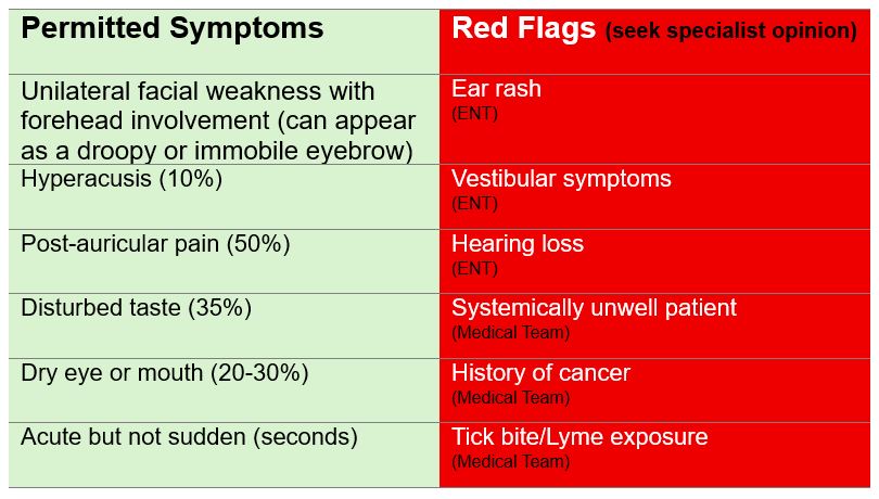
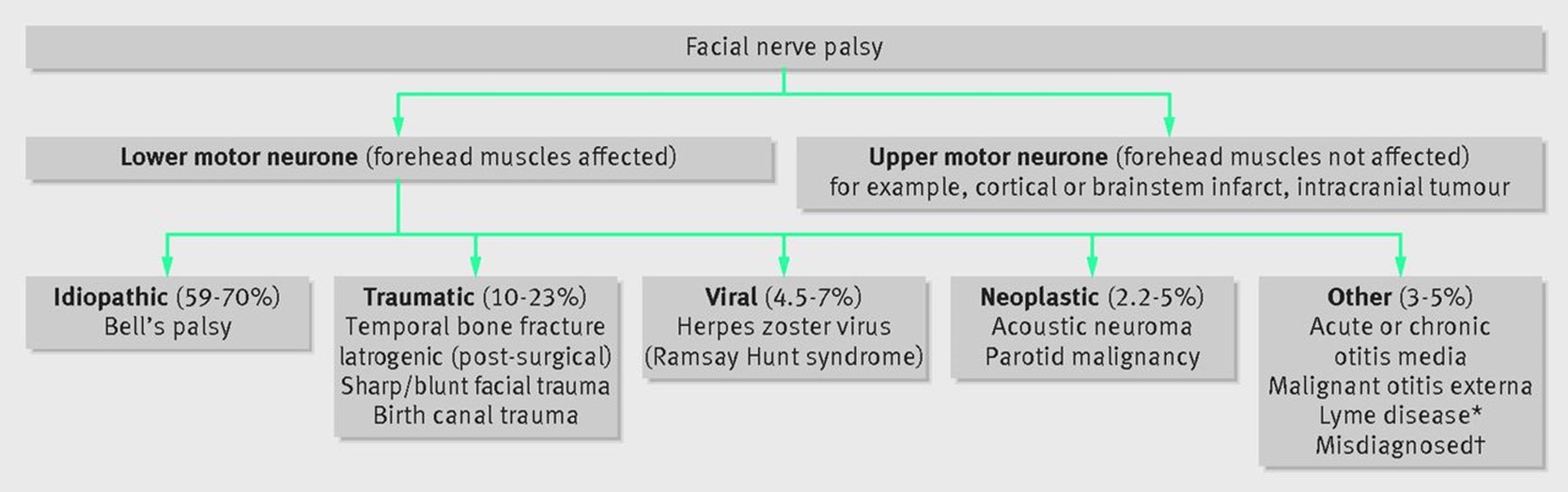
Critical differential diagnoses for isolated facial nerve palsy include:
- Stroke (CVA) or acute neurological cause such as Guillain Barre syndrome – check for other neurological symptoms and assess for forehead sparing (see below).
- Infection – assess for ENT symptoms and signs of ear canal discharge or polyp, rash. Consider Lyme disease (see separate CGO)
- Head trauma – ask about history of trauma, and look for signs of skull base or maxillofacial fracture.
- Masses in the parotid or neck - thorough examination is essential
- Ramsay Hunt (varicella zoster) - examine for vesicles around the pinna or palate, with significant pain
Note that the management of these differential diagnoses are not covered within this CGO but other guidance is available.
Audience
This guideline is intended for the use of registered healthcare professionals fulfilling a general role in forward medical locations or in an Emergency Department on deployed operations.
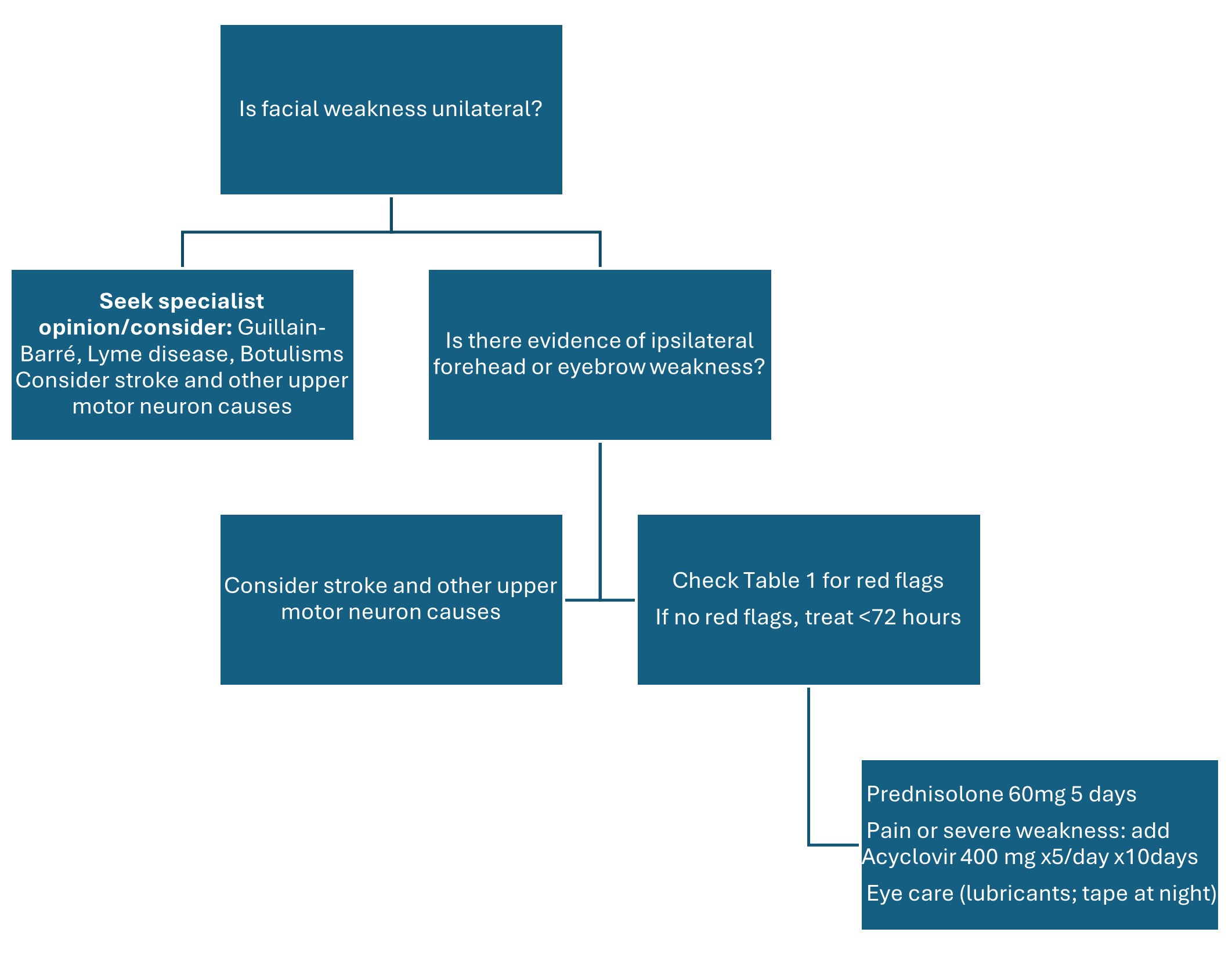
Initial Assessment & Management
Assessment
- A diagnosis of Bell's palsy can be made when no other medical condition is found to be causing facial weakness or paralysis.
- Apart from rapid onset uniformly distributed one-sided lower motor neurone facial palsy, the physical examination should be unremarkable.
- History:
- Onset and progression
- Any recent head / facial trauma or URTI
- Any previous facial palsy, face/lip/parotid swelling, uveitis
- Associated symptoms – malaise, fever, rash, headache, arthralgia, other CN issues (diplopia / hoarse voice), ear symptoms (tinnitus, hearing loss, vertigo)
- Current pregnancy
- Comorbidities / medications – immunosuppression, autoimmune disorder, neurotoxic medications, malignancy.
- Recent travel, for example to a Lyme disease endemic area
- Examination:
- Neurological examination and head and neck examination:
- Cranial nerve.
- The forehead receives bilateral upper motor neurone innervation, therefore, if there is forehead sparing in the facial palsy, it is likely to be an upper motor neurone cause (such as a stroke). A lower motor neurone cause results in weakness of the entire side of the face including the forehead.
- Ears:
- Otoscopy - Any evidence of acute / chronic otitis media or cholesteatoma.
- Weber's and Rinne's tuning fork tests (see below). Ramsay Hunt syndrome causes sensorineural hearing loss. Conductive hearing loss may indicate otitis media or cholesteatoma. Bell’s palsy may be associated with hypersensitivity to loud sounds.
- Ear canal - Malignant otitis externa can cause LMN facial nerve palsy. Vesicles may indicated Ramsay Hunt syndrome.
- Parotid gland — palpate the parotid gland and visually examine the oropharynx to rule out a deep lobe parotid tumour, which may displace the tonsil medially.
- Skin of the head, face, cheek, oral cavity, mastoid region — to check for a skin rash, nodules, or vesicles (Ramsay Hunt), ulcerative lesions, bruises (skull base fracture), swelling and tenderness (mastoiditis).
- Eyes and periocular complex.
- Peripheral nervous system exam – power, tone, coordination, reflexes, sensory examination
- Other symptoms – “bullseye” lesions suggest Lyme disease
Treatments
Steroids - If presenting within 72 hrs of onset start oral prednisolone:
- 60mg once daily for 5 days then reduce by 10mg/day (total treatment 10 days)
- Ensure PPI cover (e.g. Omeprazole, Lansoprazole) for duration of steroid course
Eye Care:
- Tape the eye closed when sleeping if the patient is unable to fully close it.
- Use regular eye lubrication (artificial tears) if available.
- Use eye protection to prevent dust / debris / sunglasses when outside.
Antivirals:
- Add Valaciclovir 1g three times daily for 7 days if paralysis is complete or almost complete (House Brackman score 5 or 6), or if vesicles are seen in the ear and/or on the palate.
General management:
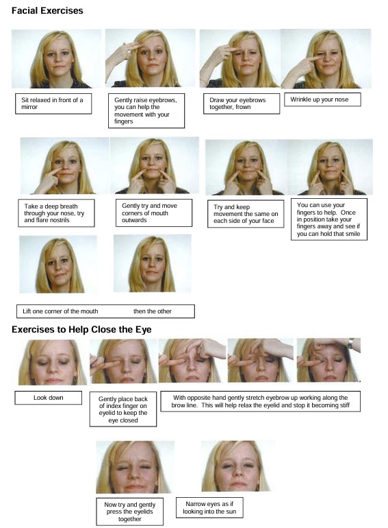
- Initiate facial nerve palsy exercises - see accordion content below.
- Advise patient to urgently seek further medical advice in the event of eye irritation / pain / itching / visual changes.
- If facial weakness affects eating – consider soft diet / use of straws if available.
Evacuate:
- Bell's Palsy typically takes weeks to resolved during which time the patient may be unable to close their eye leading to a high risk of ocular injury in the deployed environment.
- In all but exceptional situations the patient should therefore be evacuated to deployed hospital care following diagnosis.
Advanced Assessment & Management
- As per initial management.
- Consider FBC and U&E if an infective cause is suspected from clinical history and examination – fevers, systemically unwell, evidence of mastoiditis (pain and tenderness posterior to ear), vesicular lesions (Ramsay Hunt).
- Consider CT head if available in the case of diagnostic uncertainty and need to rule out upper motor neurone lesion, base of skull fracture, malignancy or mastoiditis.
Seek specialist advice if any evidence of:
- Upper motor neurone cause (limb paresis, facial paraesthesia, other cranial nerves involved, postural imbalance) – need urgent assessment as suspected stroke (see relevant CGO).
- Trauma
- Cancer (gradual onset over 6 months, pain in facial nerve distribution, lesions suggestive of cancer, ipsilateral hearing loss)
- Suspected infective cause
- Discuss with neurology / ENT if:
- Atypical features
- No improvement after 3 weeks treatment
- Changes in neurological findings
- Incomplete recover at 3 months or recurrent episodes
- Diagnosis uncertain
- Discuss with ophthalmology if any eye symptoms: pain, irritation, itch, changes in acuity.
Prolonged Casualty Care
- If evacuation is not possible, continue facial nerve palsy exercises as below.
- Wean steroids gradually.
- Ensure eye protection is carefully managed, particularly in dry austere environments.
Paediatric Considerations
The incidence of Bell's palsy in children is lower than in adults, so an especially careful assessment is needed to exclude other, potentially serious, causes of facial palsy; otherwise the management is the same as in adults.
If steroids are indicated due to presenting within 72 hours then adjust dose as per formulary.
|
House Brackmann Scoring |
|
|
1 |
Normal symmetrical facial function |
|
2 |
Slight facial weakness Eye closes Good resistance to eye opening Slight asymmetry to smile |
|
3 |
Obvious weakness Eye closes Weak resistance to eye opening Not disfiguring |
|
4 |
Obvious disfiguring weakness Unable to raise brow Incomplete eye closure and mouth asymmetry |
|
5 |
Flicker of movement only |
|
6 |
Complete paralysis |
Rinne's Test
Rinne’s test is a simple assessment comparing air conduction (AC) to bone conduction (BC) to distinguish between conductive and sensorineural hearing loss. Air conduction is normally better than bone conduction, but if bone conduction is shown to be better then this indicates conductive hearing loss.
Strike a 512 Hz tuning fork and place the base on the mastoid process (behind the ear). Ask the patient to tell you when they can no longer hear the sound.
Immediately move the fork 1–2 cm away from the ear canal (parallel to the ear) and ask if the sound is still heard.
If the sound can still be heard in front of the ear (AC) after the patient can no longer hear it behind the ear (BC), air conduction is better than bone conduction and the test result is normal.
However, if the sound is heard better, or for longer, on the mastoid bone than in front of the ear then bone conduction is better than air conduction.
Weber's Test
Weber's is a similar test that compares bone conduction in both ears. It identifies whether hearing loss is conductive (sound lateralises to the affected ear) or sensorineural (sound lateralises to the unaffected/better ear).
A tuning fork is struck and placed on the midline of the forehead.
If sound is heard equally in both ears then the result is normal.
If there is a Conductive Hearing Loss (CHL) then the sound is heard louder in (lateralises to) the affected/blocked ear.
If there is a Sensorineural Hearing Loss (SNHL) then the sound is heard louder in (lateralises to) in the unaffected/better ear.
Complementary Tests
Combining the results of both tests supports a working diagnosis pending formal audiometry and specialist assessment:
A Conductive Hearing Loss (from wax, fluid, or ear infection): will have a negative Rinne test (BC ≥ AC) in the affected ear, while Weber's test will lateralise to the affected ear.
A Sensorineural Hearing Loss (for example due to age-related hearing loss or nerve damage) will have a positive Rinne test (AC > BC) in both ears but Weber's test will lateralise to the unaffected (good) ear.