The app and website was publicly released on 2 Apr 2024
Update Jul 2026
New guidelines continue to be added.
The content will undergo continuous review by all our cadres.
Do use the search function on the app and website to make it easier to find relevant guidance.
Feedback on layout can be given below.
Saf [Updated 1 Jul 2026]
Septic Arthritis
!Warning
What's new / Latest updates
This is the first iteration of this CGO
Objectives
This guideline outlines the diagnostic considerations and procedural guidance for the management of Septic Arthritis.
The key aims are:
To ensure timely diagnosis and treatment of Septic Arthritis
To reduce morbidity by identifying and managing septic arthritis, gout, or other inflammatory conditions promptly.
To provide clear protocols for advanced assessment, prolonged casualty care, and paediatric considerations.
Scope
This guideline applies to the management of Septic Arthritis in military or deployed settings. It includes protocols for initial assessment, advanced diagnostics with the focus on differentiating the diagnosis of Septic Arthritis from other medical causes of a hot swollen joint. Management strategies of suspected Septic Arthritis, including joint aspiration and surgical wash out are outlined.
Audience
The target audience is deployed medical teams. It is particularly aimed at deployed surgical units but the material also has relevance to personnel in further forward medical locations.
Initial Assessment & Management
There is a wide-ranging differential diagnosis of an acute atraumatic monoarthritis including: gout, pseudogout and other crystal arthropathies, autoimmune causes including rheumatoid, exacerbation of osteoarthritis; bursitis; cellulitis; acute injury, Charcot joint; bone or soft tissue malignancy and septic arthritis.
History: Key features to elicit and document:
Time course of the pain and dysfunction
Any recent or preceding trauma
Previous episodes
Any known arthropathy causes
Systemic symptoms: malaise, fever, rigors
Other joints affected (if more than one joint is involved then the likelihood of a infectious cause is extremely low)
Treatment so far, particularly administration of antibiotics (document duration of treatment, name of agent, and date/time of last dose)
Examination: Key features to elicit and document:
Presence or absence of heat, erythema, swelling, tenderness
If erythema is present, draw a “tidemark” around the leading edge and annotate the date on the patient
Active and passive range of movement Distal neurovascular status Weight bearing status
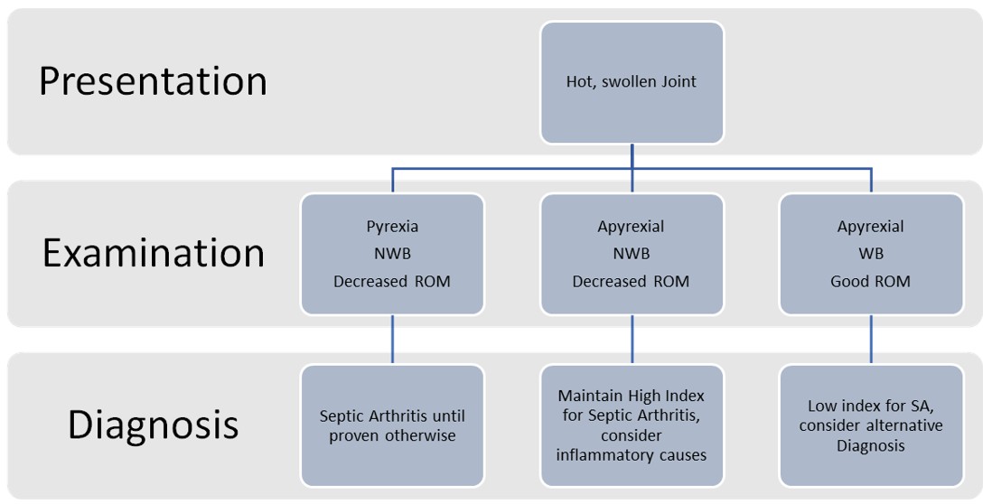
Role 1: Investigations at Role 1 will be limited. If a working diagnosis of septic arthritis is raised, the patient should be evacuated urgently to deployed orthopaedic care. If the patient is not septic, withhold antibiotics until an aspirate can be performed. If the patient is septic on presentation, they should be treated early in accordance with the CGO “Immediate Management of Septic Shock Patient” in conjunction with the deployed antimicrobial guidelines. The need for joint aspiration should not delay the administration of antibiotics in a septic patient.
Role 2: At Role 2, a hot swollen joint should be initially investigated with plain film radiograph and blood tests, which is limited to FBC with a 3-part differential. The aim of aspiration without further laboratory analysis technique is to aspirate pus as diagnostic aid opposed to guiding antimicrobial administration. CRP is only available at R2E or R3. If the echelon of care is scaled for microbiology capability, then blood cultures and a joint aspirate should be analysed.
A pragmatic approach to joint aspiration in the absence of deployed microbiological capability is as follows:
Aspirate is frank pus: a diagnosis of septic arthritis confirmed
Aspirate is turbid fluid: a diagnosis of septic arthritis confirmed
Aspirate is normal synovial fluid: consider alternative diagnosis
This approach risks over-treating hot swollen joints that may not be proven to be infected but it is the safest and most robust approach in the deployed setting.
If deployed microbiological capability allows, then the presence or absence of crystals and/or organisms can feed into the clinical decision making in the same manner as in firm base practice.
There is no role for Point of Care Ultrasound to rule out a deep joint effusion in the deployed setting.
Figure 1. Septic Arthritis Initial Management in the Deployed Setting
Advanced Assessment & Management
Joint Aspiration
In the diagnostic work up of a hot swollen joint, consideration should always be given to joint aspiration. Role 3 is likely to be the only echelon that would have the deployed capability to perform microscopy, cultures and sensitivities. Aspirate should be sent for Microscopy, Culture and Sensitivities and testing for WBC and PMN with WBC > 50,000 u/L and PMN > 90% considered strongly predictive of septic arthritis. A BMS may be able to comment on the presence of crystals, depending on their scope of practice.
Joint aspiration should be undertaken by an appropriately trained clinician, with a sterile technique and informed consent regarding the risk of introducing infection to a sterile joint. A joint aspiration should not be performed through cellulitic or broken skin.
Equipment required:
Wound pack for sterile field and sterile gloves
Cleaning solution i.e. chlorprep, alcoholic betadine
An 18 or 20 gauge needle with 10-50ml syringe
Appropriate containers i.e. universal container for microscopy and culture bottles
The landmarks for joint aspirations are noted below:
Hip: The hip is a deep joint and in UK Firm base is accessed under fluoroscopic or USS guidance. A landmark technique can be utilised based on the mid inguinal point and femoral artery, however, aspiration should be discussed with an orthopaedic surgeon.
Knee: An anterolateral or anteromedial approach is a simple approach for knee aspiration. Position the knee in extension, or slightly flexed with the patient on a stretcher. After cleaning and prepping the area, insert the needle 1-2cm lateral or medial to the proximal 1/3 of the patella into the suprapatellar pouch. Enter the skin perpendicularly and aim to go along the posterior aspect of the patella until joint aspirate is obtained.
Ankle: Position the patient supine and place a cushion under the ankle to hold it in a dorsiflexed position. Identify the medial malleolus and the tibialis anterior tendon. The insertion site is the dimple between tibialis anterior and the medial malleolus. Insert the needle perpendicular to the skin aiming towards the joint site.
Shoulder: Position the patient sitting with their arm at the side and hand on the lap across the patient. The joint is accessible posteriorly. Identify the humeral head, acromion and the coracoid process via palpation. Insert the needle in the sulcus between the acromion and humeral head, aiming toward the coracoid process anteriorly.
Elbow: Position the patient supine or sat up with the elbow flexed to 90* and the wrist pronated. Identify the radial head, ulna/olecranon process and the lateral epicondyle of the elbow. The insertion site is the depression between these three landmarks. Insert the needle perpendicular to skin and aim direct towards the antecubital fossa.
Wrist: Position the patient supine or sitting with wrist on the side. Identify lister’s tubercle of the radius (distal tubercle on dorsal radius) and identify extensor pollicis longus tendon (can be identified by extending thumb). Identify the hollow between these two structures. Apply slight traction and flex the wrist slightly. Needle entry occurs distal to lister’s tubercle and ulnar to extensor pollicis longus tendon. Aiming slightly proximally to mirror the wrists joints natural anatomy of a volar tilt. Ensure you do not insert the needle into the anatomical snuffbox (radial to EPL tendon) due to risk of neurovascular injury.
Summary of Investigations at different echelons of care:
Imaging - Plain Film
FBC
CRP
Blood Cultures
Lab Microbiology
Role 1
❌
❌
❌
❌
❌
Role 2 Fwd
✅
✅
❌
❌
❌
Role 2 Rear
✅
✅
❌
✅
✅
Role 3
✅
✅
✅
✅
✅
Table 1. Summary of Investigations available at different echelons of care
Clinical Decision-Making Tool
A web-based Septic Arthritis Risk calculator can be used to help decision-making once results from aspirate are available in difficult clinical cases. https://septicknee.org/risk-calculator
Factors to consider include synovial fluid WBC >30,000, positive synovial gram stain, synovial crystals found, history of septic arthritis in any joint, clinical effusion on exam, duration of knee pain > 2 days.
Advanced Management
This section will consider the management of Septic Arthritis. In cases where the working diagnosis is an inflammatory arthropathy or crystal arthropathy, the mainstay of treatment is analgesia and evacuation, situation dependant.
Septic Arthritis
There are three goals to management of a septic arthritis:
Source Control – achieved through aspiration and surgical washout, arthrotomy in the deployed setting
Identification and treatment of Sepsis
Minimising cartilage damage – timely decision for surgical washout.
Medical Management of Septic Arthritis:
Prompt identification of a septic patient is essential and should be managed in accordance with CGOs (Immediate Management of Septic Shock Patient). Empirical antibiotics should not be delayed for joint aspiration or surgical washout in a septic patient. In stable patients, strong consideration should be given for antibiotic administration after joint aspiration if microbiological capability is available. All patients with septic arthritis will subsequently require joint drainage; either serial aspiration or surgical drainage; whist monitoring for sepsis.
Deployed antimicrobial guidance should be followed with the "deployed secondary care cellulitis/abscesses/furunculosis" section giving the relevant agents. Typically antibiotics are given for 3-4 weeks, and an oral agent can usually be considered once clinically stable which is typically after 3-4 days once source control has been achieved. Advice can be found via the infection reach-back service available by telephone or message systems, where deployed without local military infection specialists.
Surgical Management of Septic Arthritis:
The definitive treatment for a septic arthritis is a surgical washout, although consideration could be given by the senior deployed orthopaedic surgeon for the role of aspiration to dryness.
Serial Aspirations
In situations such as prolonged evacuation timelines, or specific operational constraints, it may be appropriate to consider serial aspiration of a joint alongside intravenous antibiotics. Outcomes from serial aspiration of septic joints has been shown to be non-inferior to surgical management. However, to help guide decision making, certain parameters to predict surgery in medically managed patients have been identified including more comorbidities, a prolonged fever, and a delta CRP (increased CRP day 1 after aspiration). This approach has a high failure rate in hip and shoulder septic arthritis and should be avoided in these joints.
Whilst it is acknowledged that aspiration to dryness is not gold standard in UK Firm Base practice, within a deployed setting, clinical and tactical consideration may justify the approach. This should be discussed with a senior clinician.
Surgical Techniques - Arthrotomy
Any casualty with a suspected septic arthritis treated with an initial washout, should be reviewed for consideration of a further washout after 24-48 hrs of antibiotic treatment. The approaches for Hip, Knee, Shoulder and Ankle Arthrotomy are outlined in the accordion section below.
Drugs
Analgesia – give analgesia in accordance with guidelines and the treating clinicians scope of practice.
Antibiotics – See above regarding timing of antimicrobial therapy. Refer to the Deployed Antimicrobial Guidelines for choice of agent.
Prolonged Casualty Care
In the situation of a prolonged hold, the treatment for suspected septic arthritis should be joint aspiration for diagnosis and surgical washout with broad spectrum antibiotics. Clinicians should bear in mind the requirement for a further surgical washout in a proportion of cases.
Casualties should be closely monitored for complications such as systemic sepsis.
Casualties should be evacuated to definitive surgical care as soon as feasible.
Paediatric Considerations
In the paediatric setting, septic arthritis is a medical emergency due to the risk of growth plate damage. Early signs may be subtle - irritability, altered gait, refusal to bear weight or pseudoparalysis. Diagnostic criteria such as Kocher's criteria – weight bearing status, Temperature > 38.5°C, ESR >40mm/hr and WCC >12,000 cells/mm3 - should be used with clinical judgment to differentiate between a septic arthritis and a transient synovitis. Antibiotic treatment should be delivered as per current guidelines and is weight based. Surgical treatment of septic arthritis in the paediatric population is through surgical drainage with arthrotomy.
Surgical Techniques - Arthrotomy
Hip:
The standard approach for draining a septic hip is the anterior approach, although an operating surgeon may decide to perform a different approach based on their surgical practice and experience. The anterior approach is described: An 8-10 cm skin incision is made from the ASIS towards the lateral aspect of patella. The lateral femoral cutaneous nerve should be identified superficial to the fascia and protected by moving it medially in the wound. The fascia is incised longitudinally, and the interval between tensor fascia lata (laterally, superior gluteal nerve) and sartorius (medially, femoral nerve) is developed. The iliac origin of tensor fascia lata may be partially released to improve exposure. Deeper, the interval between rectus femoris and gluteus medius is identified and developed. The origins of rectus femoris may need to be detached in order to expose the hip capsule, which is then incised longitudinally to decompress the joint and evacuate pus.
Knee:
Arthroscopic arthrotomy:
Anterolateral: palpate lateral joint line and lateral edge of patella tendon. Leg flexed to 90 – incision, with patella tendon protected- 1-1.5cm above joint line through capsule
Anteromedial: palpate medial joint line and medial edge of patella tendon. Leg flexed to 90 – incision, with patella tendon protected- 1-1.5cm above joint line through capsule
Open Arthrotomy – Medial parapatellar
Midline skin incision centred 5cm above superior pole of patella to tibial tuberosity
Identify medial border of patella
Develop interval between VMO and medial patellofemoral retinaculum
Incision the capsule to invert patella laterally
Ankle:
Incision over anterior aspect of joint, using lateral and medial malleoli as landmarks, SPN is very superficial and should be protected. No true internervous plane – EHL and EDL define intramuscular plan. Incise fascia, identify EHL and EDL and NV bundle medial to EHL; retract EHL together with NV bundle medially EDL laterally. Continue incising down the cut through anterior capsule.
Shoulder:
The deltopectoral approach is the standard open approach for draining a septic shoulder. The incision is made along the deltopectoral groove, identifying and protecting the cephalic vein while developing the interval between deltoid (axillary nerve) and pectoralis major (medial and lateral pectoral nerves). The clavipectoral fascia is incised, and the conjoined tendon is retracted medially to expose the subscapularis. The subscapularis is incised or split close to its tendon insertion to enter the joint, allowing evacuation of pus, synovectomy, and copious lavage. Care is taken to protect the axillary nerve inferiorly and avoid damage to the long head of biceps.
Elbow:
The standard approach to drain a septic elbow is the lateral (Kocher) approach, which gives safe access to the joint while avoiding major neurovascular structures. A skin incision is made centred over the lateral epicondyle, and the interval between anconeus and extensor carpi ulnaris (ECU) is developed. Care is taken to identify and protect the posterior interosseous nerve, which lies deep and anterior to the supinator, pronation of the forearm will move the nerve further from plane of dissection. The capsule is incised longitudinally to access the ulnohumeral and radiocapitellar joints, allowing evacuation of pus, synovectomy, and irrigation. Avoid incising the capsule too far anterior as the radial nerve lie over the anterolateral portion of the elbow capsule.
Wrist:
The dorsal approach to the wrist provides safe access to the radiocarpal and midcarpal joints for washout in septic arthritis. A longitudinal incision is made just ulnar to Lister’s tubercle, centred between radia and ulnar styloids. The extensor pollicis longus (EPL) tendon is retracted radially if encountered. The extensor retinaculum is opened, and the third and fourth extensor compartments are gently retracted to expose the dorsal capsule. A longitudinal capsulotomy is performed to allow evacuation of pus, synovectomy, and copious irrigation of the radiocarpal and midcarpal joints. Care is taken to protect the dorsal sensory branches of the radial and ulnar nerves.
Last reviewed:
06/08/2026
Next review date:
06/08/2027
Version:
1
Related resources
BOAST – The Management of Children with Acute Musculoskeletal Infection
References
Rotem G, Zur-Aviran N, Sherr-Lurie N, Weltsch D, Schindler A, Tenenbaum S, et al. Treatment of Septic Arthritis of the Hip Joint Using Repeated Aspirations: When Should We Operate? Indian J Orthop 2022;56(6):1090–1095
McConnell I., Baghban A., Holleck J., Gupta S. Septic arthritis of native joints: Are outcomes better with medical or surgical management?. Open Forum Infectious Diseases 2017
Ravn C, Neyt J, Benito N, Abreu MA, Achermann Y, Bozhkova S, et al. Guideline for management of septic arthritis in native joints (SANJO). J Bone Jt Infect 2023;8(1):29–37
Flores-Robles, Bryan Josué MD*; Jiménez Palop, Mercedes MD*; et al. Medical Versus Surgical Approach to Initial Treatment in Septic Arthritis: A Single Spanish Center’s 8-Year Experience. JCR: Journal of Clinical Rheumatology 25(1):p 4-8, January 2019. | DOI: 10.1097/RHU.0000000000000615
Kocher MS, et al. Validation of a clinical prediction rule for the differentiation between septic arthritis and transient synovitis of the hip in children. JBJS. 2004; 86(8): 1629-35.
Holzmeister A, Frazzetta J, Yuan FFN, Cherones A, Summers H, Cohen J et al. Evaluation for septic arthritis of the native adult knee is aided by multivariable assessment. Am J Emerg Med. 2021 Aug;46:614-618.